Download
1 / 1
10 likes | 69 Views
A. B. 100. 20. **. **. **. **. **. **. Relative cell No. **. 50. **. % Apoptotic nuclei. 10. **. **. **. 0. 0. 5. 7. 2. 3. 4. 0. 1. 5. 5. 7. 2. 3. 4. 0. 1. 5. Days after X-rays. Days after X-rays.

E N D
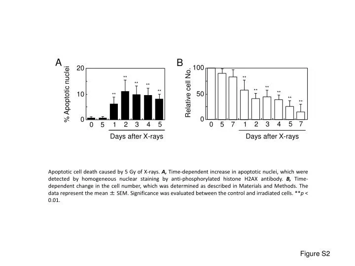
A B 100 20 ** ** ** ** ** ** Relative cell No. ** 50 ** % Apoptotic nuclei 10 ** ** ** 0 0 5 7 2 3 4 0 1 5 5 7 2 3 4 0 1 5 Days after X-rays Days after X-rays Apoptotic cell death caused by 5 Gy of X-rays. A, Time-dependent increase in apoptotic nuclei, which were detected by homogeneous nuclear staining by anti-phosphorylated histone H2AX antibody. B, Time-dependent change in the cell number, which was determined as described in Materials and Methods. The data represent the mean ± SEM. Significance was evaluated between the control and irradiated cells. **p < 0.01. Figure S2